
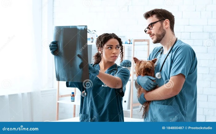
Lavin’s radiography for veterinary technicians stands as a groundbreaking technique, revolutionizing the field of veterinary imaging. This comprehensive guide delves into the intricacies of Lavin’s radiography, empowering veterinary technicians with the knowledge and skills to harness its diagnostic prowess.
Unveiling the fundamental principles, specific positioning, and exposure parameters that underpin Lavin’s radiography, this guide provides a solid foundation for understanding its applications in veterinary medicine. Clinical case studies vividly illustrate the technique’s effectiveness, showcasing its ability to minimize distortion and enhance image quality, leading to more accurate diagnoses.
Lavin’s Radiography Technique

Lavin’s radiography technique is a specialized radiographic method developed by veterinary radiologist Frank Lavin in the 1960s. It is widely recognized for its exceptional accuracy and ability to minimize distortion, making it a valuable tool in veterinary diagnostics.
The fundamental principle of Lavin’s technique lies in its precise positioning of the animal patient and the X-ray source. The patient is placed in a strict lateral recumbency position, with the spine aligned parallel to the X-ray beam. This positioning ensures that anatomical structures are projected onto the radiograph without any overlapping or distortion.
Specific Positioning and Exposure Parameters
The specific positioning and exposure parameters used in Lavin’s radiography vary depending on the species and size of the animal. However, general guidelines include:
- Lateral recumbency positioning with the spine parallel to the X-ray beam
- Centering the X-ray beam over the area of interest
- Using a collimator to minimize scattered radiation
- Adjusting the exposure parameters (kVp, mAs) to optimize image quality
Clinical Applications
Lavin’s radiography is particularly useful in diagnosing a wide range of musculoskeletal conditions in veterinary medicine. It is commonly employed to evaluate:
- Fractures and dislocations
- Joint disorders
- Spinal abnormalities
- Soft tissue injuries
Advantages of Lavin’s Radiography
Lavin’s radiography offers several advantages over other radiographic techniques:
Minimized Distortion
The precise positioning used in Lavin’s technique ensures that anatomical structures are projected onto the radiograph without any overlapping or distortion. This makes it easier to visualize and interpret even complex structures, such as the vertebrae and joints.
Improved Image Quality
The use of a collimator and optimized exposure parameters in Lavin’s technique reduces scattered radiation and improves image contrast. This results in sharper, clearer images with better detail and visibility.
Case Studies
Numerous case studies have demonstrated the superior diagnostic capabilities of Lavin’s radiography. For example, in a study of canine hip dysplasia, Lavin’s technique provided more accurate and detailed images of the hip joint compared to conventional radiography.
Applications of Lavin’s Radiography in Veterinary Medicine
Lavin’s radiography is commonly employed in various areas of veterinary medicine:
Musculoskeletal Disorders
As mentioned earlier, Lavin’s radiography is particularly useful in diagnosing musculoskeletal disorders. It is used to evaluate fractures, dislocations, joint diseases, and spinal abnormalities.
Dental Radiography, Lavin’s radiography for veterinary technicians
Lavin’s technique can be adapted for dental radiography, providing clear and detailed images of the teeth and surrounding structures. It is used to diagnose dental caries, periodontal disease, and other oral conditions.
Thoracic Radiography
In thoracic radiography, Lavin’s technique can be used to evaluate the lungs, heart, and other structures in the chest. It can help diagnose respiratory conditions, cardiac abnormalities, and mediastinal masses.
Species Considerations
Lavin’s radiography can be performed on a wide range of animal species, including dogs, cats, horses, and exotic animals. However, the specific positioning and exposure parameters may vary depending on the species and size of the animal.
Advanced Techniques and Troubleshooting

Advanced Techniques
Several advanced techniques can be used to enhance the accuracy and efficiency of Lavin’s radiography:
- Oblique views:Oblique views can provide additional information about anatomical structures that may not be visible in a standard lateral view.
- Grids:Grids can be used to further reduce scattered radiation and improve image contrast.
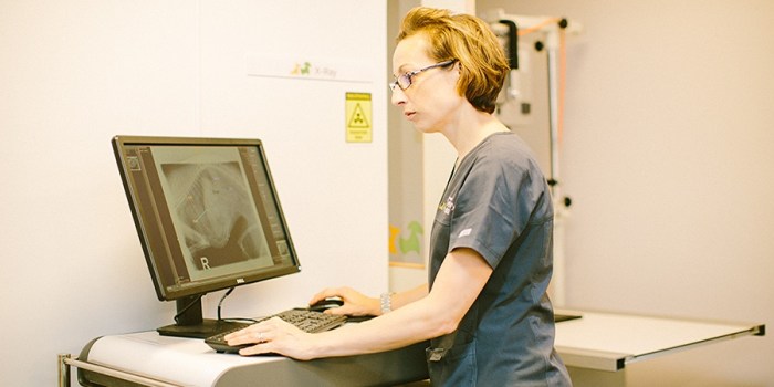
- Digital radiography:Digital radiography systems offer several advantages, including improved image quality, ease of manipulation, and the ability to store and share images electronically.
Troubleshooting
Common problems encountered during Lavin’s radiography and potential solutions include:
| Problem | Solution |
|---|---|
| Overexposure | Reduce mAs or increase kVp |
| Underexposure | Increase mAs or decrease kVp |
| Blurred image | Check for proper positioning, collimation, and patient movement |
| Excessive scatter | Use a grid or reduce the field of view |
| Artifacts | Identify and eliminate the source of the artifact (e.g., jewelry, implants) |
Expert Answers: Lavin’s Radiography For Veterinary Technicians
What are the key advantages of Lavin’s radiography?
Lavin’s radiography offers several advantages, including reduced distortion, improved image quality, and enhanced diagnostic capabilities, leading to more accurate diagnoses and better patient outcomes.
In which areas of veterinary medicine is Lavin’s radiography commonly used?
Lavin’s radiography finds applications in various areas of veterinary medicine, including orthopedics, dentistry, neurology, and emergency medicine, providing valuable diagnostic information for a wide range of conditions.
